Chilean scientists characterize the chorion of Atlantic salmon oocytes
Researchers found that the inner layer of the chorion is composed of numerous interconnected fibers forming a complex network with numerous pores of varying size.

Published
Modified
The oocytes (female germ cell) of fish are surrounded and protected by an acellular envelope called chorion which, among other functions, protects the embryo during its development against physical and chemical factors of the environment until hatching.
The chorion, an acellular envelope that surrounds the oocytes and embryos of fish, is made up of glycoproteins (choriogenins) that are incorporated in the preovulatory follicle, between the microvilli of the granulosa cells and the cytoplasmic membrane, promoting the formation of porous channels, and plays a crucial role in protecting the embryo from environmental factors until hatching.
In a new study published in the R&D section of the latest issue of Salmonexpert magazine, a medium associated with Landbased AQ, scientists from the Catholic University of Temuco and the University of La Frontera used scanning electron microscopy (SEM) to analyze the chorion of Atlantic salmon oocytes and embryos at different stages of development, and the SDS-PAGE technique to identify glycoprotein subunits associated with the chorion.
The researchers found that the inner layer of the chorion is composed of numerous interconnected fibers that form a complex network with numerous pores of varying sizes.
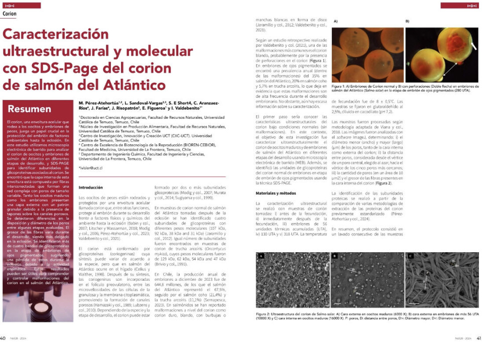
In this regard, "both mature oocytes and embryos exhibited an outer layer with a granular pattern due to the presence of plugs over the porous channels, with differences in the arrangement and diameter of the pores among some of the stages evaluated. The thickness of the fibers varies during development, being thinner at hatching," the authors explained.
On the other hand, they managed to identify more than four glycoprotein bands in the stage of pigmented eye embryos, suggesting a loss of these during hatching due to enzymatic activity.
In light of these results, the scientists concluded that the ultrastructural analysis and comparison of the protein profile of the chorion in fish, "could be a useful tool as a biomarker of early quality (mature oocytes) and/or during embryonic development".
Read the complete study in the latest edition of Salmonexpert magazine here (pages 40-45).